
Core Integration Utilizing PRI and DNS Approaches: Theoretical Constructs
Over the last several years the importance of addressing breathing patterns and training the muscles involved with respiration has gained increased attention. In parallel with this recognition, different educational institutions that focus on these structures, such as the Postural Restoration Institute (PRI) and Dynamic Neuromuscular Stabilization (DNS), have also increased in popularity. While there are applications from these two schools that are probably best suited for rehabilitation settings, there are also several direct applications to the training environment to help athletes get stronger and stay healthy.
As a brief disclaimer, my understanding of PRI is much more thorough than DNS. I've now taken 4 PRI courses (one of them twice), as well as gone through the PRT credentialing process; with regards to DNS, I've only completed the ‘A' course. The thoughts below are based strictly on my interpretations of the information I've had access to, and don't necessarily reflect the original intent of these educational resources.
Concept 1: Position Drives Function
In order for a muscle to function properly, it needs to be positioned properly. PRI uses the word “neutrality” where DNS uses “centrated”, but the underlying thought process is the same. If a joint does not possess the ability to produce full reciprocal movement (e.g. the ability to get into and out of joint-specific extreme ROMs freely), the feedback to the central nervous system (CNS) will alter the function of the surrounding muscles and movement patterns as a whole. One easy way to visualize this concept is to think of Charlie Weingroff's “Core Pendulum Theory”. Using the image below as a guide, the central nervous system knows to center a joint at “3” because it's aware that it has the ability to freely move into “1” and “5”. If something limited ROM such that the joint could no longer reach “5”, the joint may “recentrate” in a position somewhere between “2” and “3”. While this may be overly simplistic, it provides a conceptual framework from which to understand this interaction.
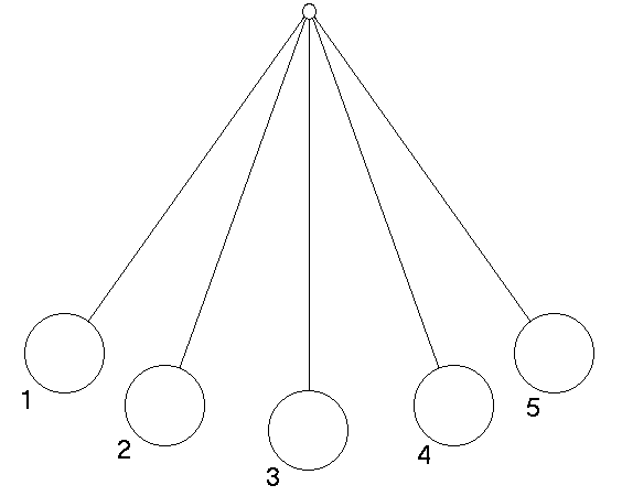
Pendulums: A visual construct of the influence of joint centration on proprioception.
Naturally, limitations in joint ROM also heavily influence muscle position and function. If, as a common example, the pelvis as a whole is rotated toward the right in a manner such that the left hemipelvis is positioned in a state of forward rotation and anterior tilt, and the right hemipelvis is in a position of backward rotation and posterior tilt, the extension fibers of the glute max will be shortened and potentially over-facilitated on the right (secondary to position of extension), and long and weak on the left (secondary to position of flexion); on the contrary, the external rotation fibers will be short and over-facilitated on the left (secondary to compensatory external rotation of femur within an acetabulum that is rotated forward), and long and weak on the right (secondary to compensatory internal rotation of the femur within an acetabulum that is rotated backward). This position also frequently leads to laxity in the left anterior hip capsule, which is a common underlying cause of groin pain in hockey players.
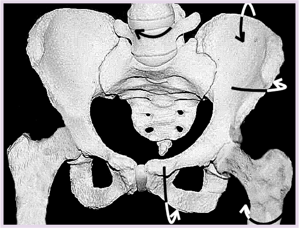
A common skeletal position of the pelvis, femur, and lumbar spine with arrows to help guide your attention. Note the asymmetry in the space surrounding the sacrum, the heights of the ASIS's, and the positioning of the obturator foramen. Anterior View.
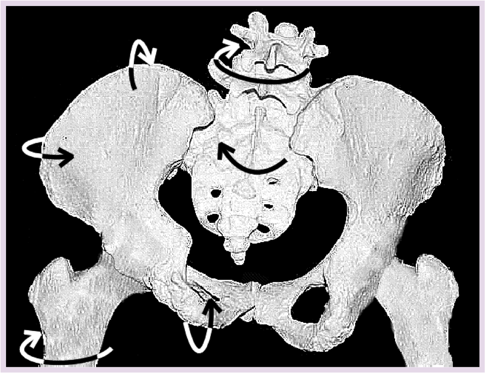
Posterior View
Images reprinted with permission from the Postural Restoration Institute®
Within this position, it can be expected that the various tensional receptors within the muscle, tendons, capsules, and joints will shift their sensitivities based on whether the structure is short or long. For example, muscle spindles, which are known to detect changes in muscle length at all lengths, but are more active at increasing lengths, will essentially down-regulate their sensitivity at any given length and shift it to longer lengths to accommodate the ongoing stretch signal created by the positional demand. Naturally, this can have a profound influence on joint proprioception, muscle firing patterns, and movement patterns in general.
In this environment, strategies to add length to a structure, range of motion to a joint, or strength to a muscle/pattern should be designed with an understanding of the joint's current position of neutrality. In the above example, restoration of a neutral pelvic alignment will reposition the surrounding muscles and open a window to reinforce this new position through active muscular control (typically asymmetrically, given the different roles the muscles play based on reversing the aforementioned compensations). Approaching asymmetrical exercise prior to re-establishing a neutral starting position will simply reinforce the compensatory pattern.
Potentially a more easily understood reference lies in a common finding in the Functional Movement Screen's Active Straight Leg Raise test. An anteriorly tipped pelvis, which is more commonly found on the left, will place the hamstrings on a stretch, limiting ASLR motion on that side relative to the right. Sometimes this limitation falls within a range that causes the score to be different from the right; sometimes it doesn't, which may be an underlying reason in why more people don't notice this asymmetry. As the FMS team makes a strong effort to mention in their courses, there are several reasons why an ASLR may be limited, and hamstring length is only one of them. Nonetheless, the most direct strategy in this situation would be to restore a neutral pelvic alignment, which would take the left hamstring off stretch and restore a symmetrical ASLR.
Concept 2: The Diaphragm Drives Everything
The diaphragm may be the single most important and underappreciated muscle within the body, playing a huge role in both respiration and core stability.
Upon inhalation, the diaphragm should descend inferiorly, creating a larger area for the lungs to expand. Upon exhalation, the diaphragm should ascend superiorly back to its starting position. Concomitant with these movements, the pelvic floor musculature, which collectively is shaped like a bowl, should be in synchrony in the same direction with the diaphragm, descending upon inhalation and ascending upon exhalation. It's this synchronous movement that helps facilitate intra-abdominal pressure and pelvic and spinal stability. This basically means that on inhales, the abdominals relax and on exhales the abdominals contract.
Likewise, the above diaphragmatic movement pattern is essential for proper air exchange. A dysfunctional breathing pattern, (e.g. a more chest-dominant pattern) can cause “hyperinflation”, which is related to a cascade of undesirable events including an increase in the body's pH and an increase in sympathetic tone of the autonomic nervous system, with all the associated consequences of this shift (e.g. increased anxiousness, sensitivity to sound/light, upregulated muscle tone, etc.). The immediacy of this relationship is highlighted by a quote pulled from the PRI Advanced Integration binder:
“We can easily disrupt this (pH) balance with 2 or 3 deep breaths. pH will rise from 7.4 to 7.5 or CO2 will fall from a normal 40 to 30 or 25 (mmHg) in less than 30 seconds (with thoracic breathing).” Gilbert, C. Journal of Bodywork and Movement Therapy, July 98.
It is these consequences that really highlight the importance of addressing breathing patterns. If an athlete relies too heavily on this chest or “thoracic” breathing strategy, they'll essentially be living in a sympathetic state, which has significant implications on the stress-load to their body, and, naturally, their ability to recover from sport- and training-related stresses.
In light of the initial discussion on position, a postural position of excessive extension through the lower thoracic and lumbar spine will place the diaphragm in a more horizontal position. This position, which is extremely common in most athletic populations, decreases the height between the peak of the diaphragm dome and it's inferior attachment points along the anterior rib cage, also known as the “zone of apposition”. When this occurs, unilaterally or bilaterally, the diaphragm on the affected side(s) up-regulates its role as a postural muscle and down-regulates its role as a respiratory muscle, which both negatively affects air exchange and core stability.
Furthermore, an excessive lower thoracic and lumbar extension is typically coupled with excessive anterior pelvic tilt. In this situation, the inverted bowl of the diaphragm and the bowl of the pelvic floor are no longer stacked and cannot function in a piston-like manner. Instead, the two are oriented in what DNS would describe as an “open-scissor” position, angling toward one another posteriorly and away from one another anteriorly.
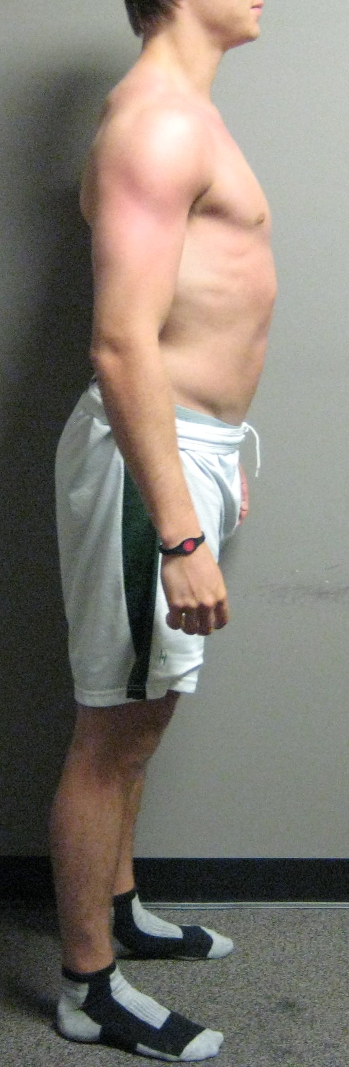
Demonstration of an open scissors posture that is extremely common in ice hockey players. Note the anteriorly tipped pelvis (look for iliac crest) and rib flare.
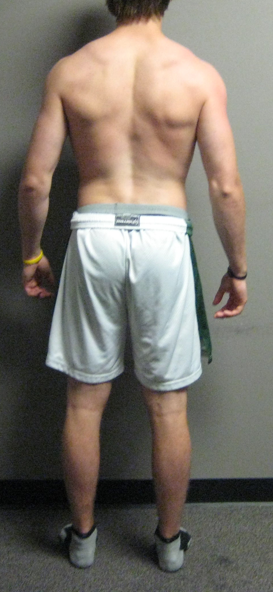
Open scissors posture: posterior view. Note the clear crease in the upper lumbar region. You can also gain an appreciation for the rotational and asymmetrical nature of this individual's pattern. Note that his pelvis is rotated to the right (visible green line on shorts on right, not on left), and that his scapula is depressed (acromion and hand visibly lower on right) and anteriorly tipped (visible inferior angle). You may also note the influence this position has on the humerus, as the left hand is clearly turned back toward the camera (internally rotated) more than the right.
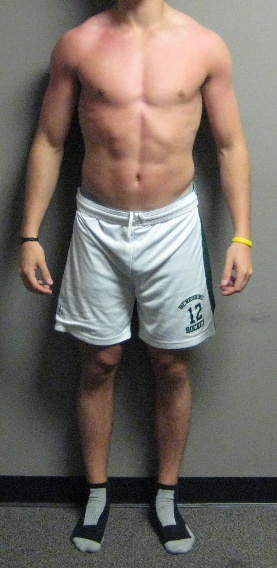
Anterior view. This angle makes it easy to confirm the right pelvic rotation we saw from behind as well as the anterior scapular tilt on the right. Also note the bilateral rib flare, which is clearly worse on the left, and that the left pec looks visibly more stretched than the right. These are all extremely common findings.
This posture will limit posterior expansion and promote excessive anterior movement of the abdomen with inhalation. While people often describe optimal diaphragmatic breathing as “belly breathing”; in reality, the goal is circumferential expansion, which is associated with a lateral expansion of the lower ribs. Dysfunctional “belly breathing” can result in abdominal dystonia, and ultimately is a symptom of lost intra-abdominal pressure and compromised core stability. Restoring an optimal spinal and rib cage alignment is a central tenant in both PRI and DNS philosophies, and is the key to creating spinal stabilization and force transfer between the lower and upper extremities.
Part 2 of this series will expand on the above constructs with practical examples of how to make mild adjustments to your current training programs to put a greater emphasis on proper alignment and function of the diaphragm.